
Page 318 - Atlas of Small Animal CT and MRI
P. 318
308 Atlas of Small Animal CT and MRI
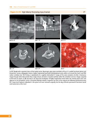
Figure 3.2.14 High‐Velocity Penetrating Injury (Canine) CT
(a) DX, LAT (b) CT, SP (c) CT, TP
(d) CT, TP (e) CT, TP (f) GP
2y MC Beagle with a gunshot injury of the lumbar spine. Neurologic signs were consistent with an L4–caudal functional spinal cord
transection. Survey radiographs reveal a highly fragmented lead bullet distributed primarily within and around the fourth and fifth
lumbar vertebrae (a). This finding is reproduced on a sagittal reformatted CT image of the same anatomic site (b). A transverse
image through mid L4 documents that a large volume of metal is located centrally within the vertebral canal (c). Images acquired
at the level of cranial L5 (d) and mid L5 (e) show the distribution of highly fragmented metal within the ventral spinal canal and
adjacent to the left pedicle, which is fractured. Multiple metallic fragments are seen in the surgical site following hemilaminectomy
(f). A large lead fragment embedded in the spinal cord was removed surgically, but the dog suffered a permanent functional spinal
cord transection at this level.
308