
Page 317 - Atlas of Small Animal CT and MRI
P. 317
Traumatic and Vascular Disorders 307
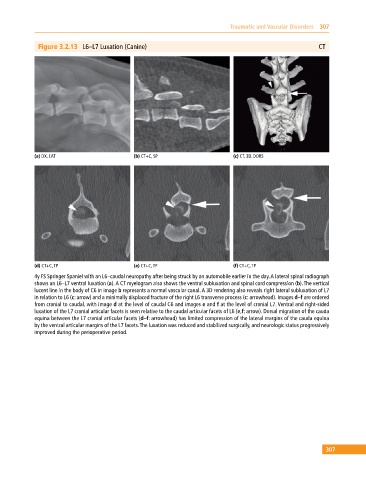
Figure 3.2.13 L6–L7 Luxation (Canine) CT
(a) DX, LAT (b) CT+C, SP (c) CT, 3D, DORS
(d) CT+C, TP (e) CT+C, TP (f) CT+C, TP
4y FS Springer Spaniel with an L6–caudal neuropathy after being struck by an automobile earlier in the day. A lateral spinal radiograph
shows an L6–L7 ventral luxation (a). A CT myelogram also shows the ventral subluxation and spinal cord compression (b). The vertical
lucent line in the body of C6 in image b represents a normal vascular canal. A 3D rendering also reveals right lateral subluxation of L7
in relation to L6 (c: arrow) and a minimally displaced fracture of the right L6 transverse process (c: arrowhead). Images d–f are ordered
from cranial to caudal, with image d at the level of caudal C6 and images e and f at the level of cranial L7. Ventral and right‐sided
luxation of the L7 cranial articular facets is seen relative to the caudal articular facets of L6 (e,f: arrow). Dorsal migration of the cauda
equina between the L7 cranial articular facets (d–f: arrowhead) has limited compression of the lateral margins of the cauda equina
by the ventral articular margins of the L7 facets. The luxation was reduced and stabilized surgically, and neurologic status progressively
improved during the perioperative period.
307