
Page 314 - Atlas of Small Animal CT and MRI
P. 314
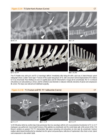
Figure 3.2.9  T5 Salter-Harris Fracture (Canine)                                             CT


             (a) DX, LAT                                                       (b) CT, SP


             (c) CT, 3D, LEFT                                                  (d) CT, 3D, OBL
             7mo FS Poodle cross with pain and T3–L3 neurologic deficits immediately after being hit with a pick‐axe. A lateral thoracic spinal
             radiograph shows a Salter–Harris type 1 fracture of the caudal body physis of T5, with concurrent ventral displacement of T6 relative
             to T5 (a: arrowhead). These findings are seen on sagittal plane and 3D reformatted CT images (b–d: arrowheads). The T5–6 vertebral
             articulations are subluxated (a,c: arrow), and the vertebral canal diameter is reduced, indirectly indicative of spinal cord compression
             (b: arrow). The injury was reduced and stabilized surgically.


              Figure 3.2.10  T12 Fracture and T10–T11 Subluxation (Canine)                                 CT


             (a) CT+C, TP                     (b) CT+C, SP                     (c) CT+C, TP
             1y M Chihuahua bitten by another dog 6 days previously. Now has neurologic deficits with neuroanatomical localization to T3–L3. A CT
             myelogram was performed. Comminuted fractures of the pedicles (a: arrowheads) and the cranial body (a,b: white arrow) of the 11th
             thoracic vertebra are present. T10–T11 intervertebral disk space narrowing and subluxation are also seen (b: arrowhead). Contrast
             medium administered intrathecally has absorbed into the spinal cord parenchyma, indicative of myelomalacia (b,c: black arrow). Spinal
             cord compression is also evident (a,b).