
Page 310 - Atlas of Small Animal CT and MRI
P. 310
300 Atlas of Small Animal CT and MRI
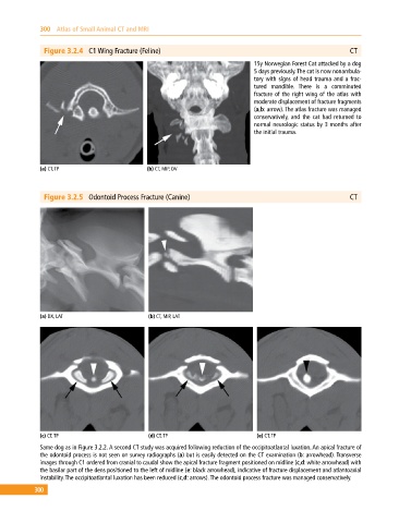
Figure 3.2.4 C1 Wing Fracture (Feline) CT
15y Norwegian Forest Cat attacked by a dog
5 days previously. The cat is now nonambula-
tory with signs of head trauma and a frac-
tured mandible. There is a comminuted
fracture of the right wing of the atlas with
moderate displacement of fracture fragments
(a,b: arrow). The atlas fracture was managed
conservatively, and the cat had returned to
normal neurologic status by 3 months after
the initial trauma.
(a) CT, TP (b) CT, MIP, DV
Figure 3.2.5 Odontoid Process Fracture (Canine) CT
(a) DX, LAT (b) CT, MIP, LAT
(c) CT, TP (d) CT, TP (e) CT, TP
Same dog as in Figure 3.2.2. A second CT study was acquired following reduction of the occipitoatlantal luxation. An apical fracture of
the odontoid process is not seen on survey radiographs (a) but is easily detected on the CT examination (b: arrowhead). Transverse
images through C1 ordered from cranial to caudal show the apical fracture fragment positioned on midline (c,d: white arrowhead) with
the basilar part of the dens positioned to the left of midline (e: black arrowhead), indicative of fracture displacement and atlantoaxial
instability. The occipitoatlantal luxation has been reduced (c,d: arrows). The odontoid process fracture was managed conservatively.
300