
Page 313 - Atlas of Small Animal CT and MRI
P. 313
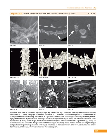
Traumatic and Vascular Disorders  303


              Figure 3.2.8  Cervical Vertebral Subluxation with Articular Facet Fracture (Canine)    CT & MR


            (a) DX, LAT                      (b) CT, SP                       (c) CT, TP


            (d) CT, 3D, DORS                 (e) CT, 3D, RLAT                 (f) CT, 3D, LLAT


            (g) T1+C, TP                     (h) T2, TP                       (i) T2, SP
            5y F Terrier cross bitten in the cervical region by a larger dog earlier in the day. Currently has neurologic deficits neuroanatomically
            localized to C6–T2. Survey radiographs reveal dorsal subluxation of C7 relative to C6 and narrowing of the C6–7 intervertebral disk
            space (a: arrowhead). Similar findings are also seen on sagittal and 3D reformatted CT images (b,f: arrowhead). In addition, there is a
            highly comminuted and displaced fracture of the right cranial articular process of C7 (c–e: arrow). The left articular process is normal
            by comparison (f: arrow). On MR images, the C6–7 intervertebral disk space is reduced in width and T2 signal intensity (i: arrow), and
            extruded disk material is present in the right ventral vertebral canal (g,h: arrowhead). There is evidence of spinal cord compression (g–i)
            and spinal cord T2 hyperintensity at the level of C6–7 (h,i), indicative of an intrinsic component to the injury. Disk material was removed
            from the canal, and the subluxation was reduced and stabilized surgically.

                                                                                                             303