
Page 309 - Atlas of Small Animal CT and MRI
P. 309
Traumatic and Vascular Disorders 299
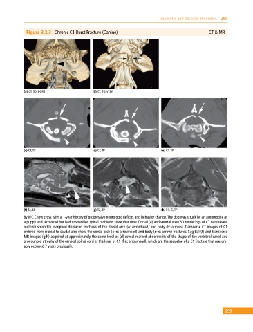
Figure 3.2.3 Chronic C1 Burst Fracture (Canine) CT & MR
(a) CT, 3D, DORS (b) CT, 3D, VENT
(c) CT, TP (d) CT, TP (e) CT, TP
(f) T2, SP (g) T2, TP (h) T1+C, TP
8y MC Chow cross with a 1‐year history of progressive neurologic deficits and behavior change. The dog was struck by an automobile as
a puppy and recovered but had unspecified spinal problems since that time. Dorsal (a) and ventral view 3D renderings of CT data reveal
multiple smoothly margined displaced fractures of the dorsal arch (a: arrowhead) and body (b: arrows). Transverse CT images of C1
ordered from cranial to caudal also show the dorsal arch (c–e: arrowhead) and body (c–e: arrow) fractures. Sagittal (f) and transverse
MR images (g,h) acquired at approximately the same level as (d) reveal marked abnormality of the shape of the vertebral canal and
pronounced atrophy of the cervical spinal cord at the level of C1 (f,g: arrowhead), which are the sequelae of a C1 fracture that presum-
ably occurred 7 years previously.
299