
Page 315 - Atlas of Small Animal CT and MRI
P. 315
Traumatic and Vascular Disorders 305
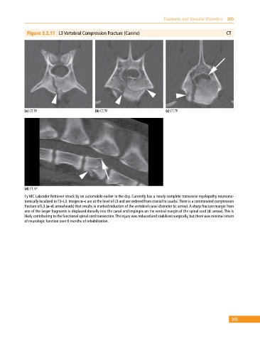
Figure 3.2.11 L3 Vertebral Compression Fracture (Canine) CT
(a) CT, TP (b) CT, TP (c) CT, TP
(d) CT, SP
1y MC Labrador Retriever struck by an automobile earlier in the day. Currently has a nearly complete transverse myelopathy neuroana-
tomically localized to T3–L3. Images a–c are at the level of L3 and are ordered from cranial to caudal. There is a comminuted compression
fracture of L3 (a–d: arrowheads) that results in marked reduction of the vertebral canal diameter (c: arrow). A sharp fracture margin from
one of the larger fragments is displaced dorsally into the canal and impinges on the ventral margin of the spinal cord (d: arrow). This is
likely contributing to the functional spinal cord transection. The injury was reduced and stabilized surgically, but there was minimal return
of neurologic function over 6 months of rehabilitation.
305