
Page 324 - Atlas of Small Animal CT and MRI
P. 324
314 Atlas of Small Animal CT and MRI
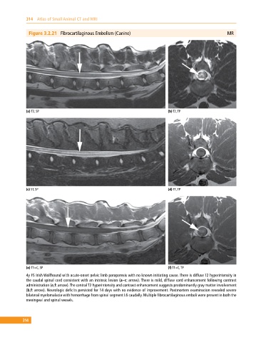
Figure 3.2.21 Fibrocartilaginous Embolism (Canine) MR
(a) T2, SP (b) T2, TP
(c) ST, SP (d) T1, TP
(e) T1+C, SP (f) T1+C, TP
4y FS Irish Wolfhound with acute‐onset pelvic limb paraparesis with no known initiating cause. There is diffuse T2 hyperintensity in
the caudal spinal cord consistent with an intrinsic lesion (a–c: arrow). There is mild, diffuse cord enhancement following contrast
administration (e,f: arrow). The central T2 hyperintensity and contrast enhancement suggests predominantly gray matter involvement
(b,f: arrow). Neurologic deficits persisted for 14 days with no evidence of improvement. Postmortem examination revealed severe
bilateral myelomalacia with hemorrhage from spinal segment L6 caudally. Multiple fibrocartilaginous emboli were present in both the
meningeal and spinal vessels.
314