
Page 329 - Atlas of Small Animal CT and MRI
P. 329
Inflammatory Disorders 319
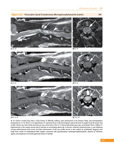
Figure 3.3.1 Presumptive Spinal Granulomatous Meningoencephalomyelitis (Canine) MR
(a) T2, SP (b) T2, TP
(c) T1, SP (d) T1, TP
(e) T1+C, SP (f) T1+C, TP
4y FS Chinese Crested Dog with a 3‐day history of difficulty walking, most pronounced in the thoracic limbs, and neuroanatomic
localization to C1–T2. There is a T2 hypointense, T1 isointense focus in the dorsal spinal cord at the level of caudal C2 (a–d: arrow). Two
smaller intrinsic foci with similar intensity are seen at the level of the C2–3 intervertebral disk space (a,c: arrowhead). There is diffuse T2
hyperintensity in the cranial cervical spinal cord due to surrounding edema (a). A discrete, uniformly enhancing mass is seen following
contrast administration (e,f: arrow), and faint enhancement of the two smaller lesions is also evident (e: arrowhead). Diagnosis was
made from results of cerebrospinal fluid analysis consistent with granulomatous meningoencephalomyelitis, absence of infectious
agents, and response to immunosuppressive doses of steroids.
319