
Page 333 - Atlas of Small Animal CT and MRI
P. 333
Inflammatory Disorders 323
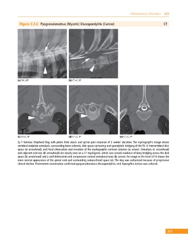
Figure 3.3.6 Pyogranulomatous (Mycotic) Discospondylitis (Canine) CT
(a) DX, LAT (b) CT+C, SP
(c) CT+C, TP (d) CT+C, TP (e) CT+C, TP
2y F German Shepherd Dog with pelvic limb ataxia and spinal pain response of 2 weeks’ duration. The myelographic image shows
vertebral endplate osteolysis, surrounding bone sclerosis, disk space narrowing and spondylotic bridging of the T5–6 intervertebral disk
space (a: arrowhead), and focal attenuation and elevation of the myelographic contrast columns (a: arrow). Osteolysis (c: arrowhead)
and adjacent sclerosis (d: arrowhead) are clearly seen on a CT myelogram, which also reveals evidence of bony bridging across the disk
space (b: arrowhead) and a well‐delineated cord compressive ventral extradural mass (b: arrow). An image at the level of T4 shows the
more normal appearance of the spinal cord and surrounding subarachnoid space (e). The dog was euthanized because of progressive
clinical decline. Postmortem examination confirmed pyogranulomatous discospondylitis, and Aspergillus terreus was cultured.
323