
Page 337 - Atlas of Small Animal CT and MRI
P. 337
Inflammatory Disorders 327
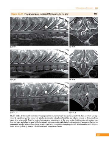
Figure 3.3.11 Pyogranulomatous (Amoebic) Meningomyelitis (Canine) MR
(a) T2, SP (b) T1, SP
(c) T1, SP (d) T1, TP
(e) T1+C, SP (f) T1+C, TP
11y MC Golden Retriever with recent‐onset neurologic deficits neuroanatomically localized between T3–L3. There is intrinsic heteroge
neous T2 hyperintensity of the midthoracic spinal cord associated with a loss of definition and reducing intensity of the subarachnoid
space (a,b: arrowheads). There is marked, heterogeneous enhancement in the same region following contrast administration
(e: arrowheads), and enhancing material fills the subarachnoid space, causing marked spinal cord compression (f: arrowhead). Postmortem
examination revealed severe, diffuse, chronic necrotizing and pyogranulomatous meningomyelitis with Acanthamoeba cysts and tropho
zoites. Neurologic findings were part of more widespread multisystem infection.
327