
Page 335 - Atlas of Small Animal CT and MRI
P. 335
Inflammatory Disorders 325
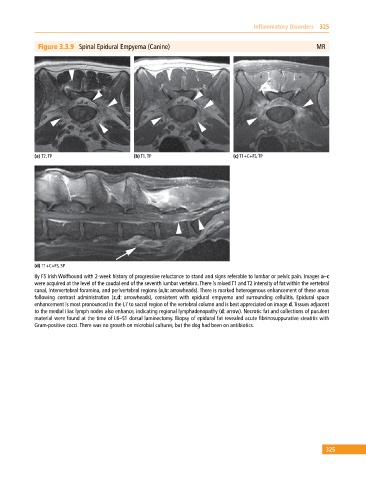
Figure 3.3.9 Spinal Epidural Empyema (Canine) MR
(a) T2, TP (b) T1, TP (c) T1+C+FS, TP
(d) T1+C+FS, SP
8y FS Irish Wolfhound with 2‐week history of progressive reluctance to stand and signs referable to lumbar or pelvic pain. Images a–c
were acquired at the level of the caudal end of the seventh lumbar vertebra. There is mixed T1 and T2 intensity of fat within the vertebral
canal, intervertebral foramina, and perivertebral regions (a,b: arrowheads). There is marked heterogenous enhancement of these areas
following contrast administration (c,d: arrowheads), consistent with epidural empyema and surrounding cellulitis. Epidural space
enhancement is most pronounced in the L7 to sacral region of the vertebral column and is best appreciated on image d. Tissues adjacent
to the medial iliac lymph nodes also enhance, indicating regional lymphadenopathy (d: arrow). Necrotic fat and collections of purulent
material were found at the time of L6–S1 dorsal laminectomy. Biopsy of epidural fat revealed acute fibrinosuppurative steatitis with
Gram‐positive cocci. There was no growth on microbial cultures, but the dog had been on antibiotics.
325