
Page 330 - Atlas of Small Animal CT and MRI
P. 330
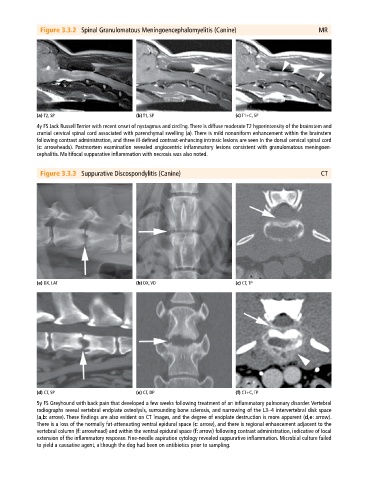
Figure 3.3.2 Spinal Granulomatous Meningoencephalomyelitis (Canine) MR
(a) T2, SP (b) T1, SP (c) T1+C, SP
4y FS Jack Russell Terrier with recent onset of nystagmus and circling. There is diffuse moderate T2 hyperintensity of the brainstem and
cranial cervical spinal cord associated with parenchymal swelling (a). There is mild nonuniform enhancement within the brainstem
following contrast administration, and three ill‐defined contrast‐enhancing intrinsic lesions are seen in the dorsal cervical spinal cord
(c: arrowheads). Postmortem examination revealed angiocentric inflammatory lesions consistent with granulomatous meningoen
cephalitis. Multifocal suppurative inflammation with necrosis was also noted.
Figure 3.3.3 Suppurative Discospondylitis (Canine) CT
(a) DX, LAT (b) DX, VD (c) CT, TP
(d) CT, SP (e) CT, DP (f) CT+C, TP
5y FS Greyhound with back pain that developed a few weeks following treatment of an inflammatory pulmonary disorder. Vertebral
radiographs reveal vertebral endplate osteolysis, surrounding bone sclerosis, and narrowing of the L3–4 intervertebral disk space
(a,b: arrow). These findings are also evident on CT images, and the degree of endplate destruction is more apparent (d,e: arrow).
There is a loss of the normally fat‐attenuating ventral epidural space (c: arrow), and there is regional enhancement adjacent to the
vertebral column (f: arrowhead) and within the ventral epidural space (f: arrow) following contrast administration, indicative of local
extension of the inflammatory response. Fine‐needle aspiration cytology revealed suppurative inflammation. Microbial culture failed
to yield a causative agent, although the dog had been on antibiotics prior to sampling.