
Page 325 - Atlas of Small Animal CT and MRI
P. 325
Traumatic and Vascular Disorders 315
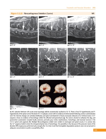
Figure 3.2.22 Fibrocartilagenous Embolism (Canine) MR
(a) T2, SP (b) T1, SP (c) T1+C, SP
(d) T2, TP (e) T1, TP (f) T1+C, TP
(g) T2*, TP (i) GP, TP
8y MC Labrador Retriever with acute‐onset neurologic deficits anatomically localized to C6–T2. There is focal T2 hyperintensity and T1
hypointensity of the spinal cord at the level of C5–6 (a,b,d,e: arrow) with no evidence of enhancement following contrast administration
(c,f). The intensity changes are centrally distributed, and spinal cord diameter is focally increased, indicative of an intrinsic lesion. A T2*
sequence shows no evidence of hemorrhage within the affected cord parenchyma (g). The owners elected to euthanize the dog, and
postmortem examination revealed locally extensive, primarily gray matter, myelomalacia, hemorrhage, and neuronal necrosis in the
C5–C7 spinal cord segments (h). Multiple fibrocartilaginous emboli were evident in meningeal and spinal vessels. It is unclear why
the T2* sequence failed to show the intraparenchymal sites of hemorrhage. One possibility is that because of the peracute nature of the
disorder, MR imaging was performed early enough after the initial insult that hemoglobin degradation products were not yet present in
adequate concentration to yield a susceptibility effect.
315