
Page 334 - Atlas of Small Animal CT and MRI
P. 334
324 Atlas of Small Animal CT and MRI
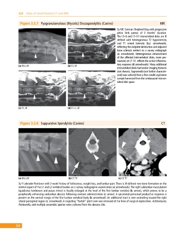
Figure 3.3.7 Pyogranulomatous (Mycotic) Discospondylitis (Canine) MR
5y MC German Shepherd Dog with progressive
pelvic limb paresis of 3 months’ duration.
The L5–6 and L7–S1 intervertebral disks are ill
defined with heterogeneous T2 hypointensity
and T1 mixed intensity (b,c: arrowheads),
reflecting the endplate destruction and adjacent
bone sclerosis evident on a survey radiograph
(a: arrowheads). Heterogeneous enhancement
of the affected intervertebral disks, more pro
nounced at L7–S1, reflects the active inflamma
tory response (d: arrowheads). Many additional
(a) DX, LAT (b) T2, SP
intervertebral disks had similar imaging features
(not shown). Sagnomella (not further character
ized) was cultured from a fine‐needle aspiration
sample harvested from the lumbosacral interver
tebral disk space.
(c) T1, SP (d) T1+C, SP
Figure 3.3.8 Suppurative Spondylitis (Canine) CT
(a) DX, LAT (b) CT, TP (c) CT, TP
3y F Labrador Retriever with 2‐week history of listlessness, weight loss, and lumbar pain. There is ill‐defined new bone formation on the
ventral aspect of the L1 and L2 vertebral bodies on a survey radiographic examination (a: arrowheads). The right sublumbar musculature
(quadratus lumborum and psoas minor) is focally enlarged at the level of the first lumbar vertebra (b: arrow), which proves to be a
peripherally enhancing sublumbar abscess following contrast administration (c: arrow). A spiculated periosteal productive response is
present on the ventral margin of the first lumbar vertebral body (b: arrowhead). An additional tract is seen extending toward the right
lateral paraspinal region (c: arrowhead). A migrating “foxtail” plant awn was removed at the time of surgical exploration. Actinomyces,
Pasteurella, and multiple anaerobic species were cultured from the abscess site.
324