
Page 336 - Atlas of Small Animal CT and MRI
P. 336
326 Atlas of Small Animal CT and MRI
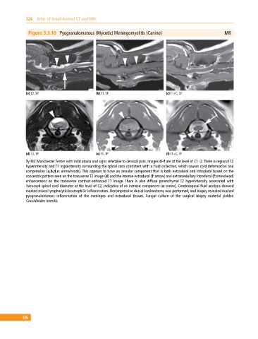
Figure 3.3.10 Pyogranulomatous (Mycotic) Meningomyelitis (Canine) MR
(a) T2, SP (b) T1, SP (c) T1+C, SP
(d) T2, TP (e) T1, TP (f) T1+C, TP
9y MC Manchester Terrier with mild ataxia and signs referable to cervical pain. Images d–f are at the level of C1–2. There is regional T2
hyperintensity and T1 hypointensity surrounding the spinal cord consistent with a fluid collection, which causes cord deformation and
compression (a,b,d,e: arrowheads). This appears to have an annular component that is both extradural and intradural based on the
concentric pattern seen on the transverse T2 image (d) and the intense extradural (f: arrow) and extramedullary intradural (f:arrowhead)
enhancement on the transverse contrast‐enhanced T1 image. There is also diffuse parenchymal T2 hyperintensity associated with
increased spinal cord diameter at the level of C2, indicative of an intrinsic component (a: arrow). Cerebrospinal fluid analysis showed
marked mixed lymphocytic/neutrophilic inflammation. Decompressive dorsal laminectomy was performed, and biopsy revealed marked
pyogranulomatous inflammation of the meninges and extradural tissues. Fungal culture of the surgical biopsy material yielded
Coccidioides immitis.
326