
Page 342 - Atlas of Small Animal CT and MRI
P. 342
332 Atlas of Small Animal CT and MRI
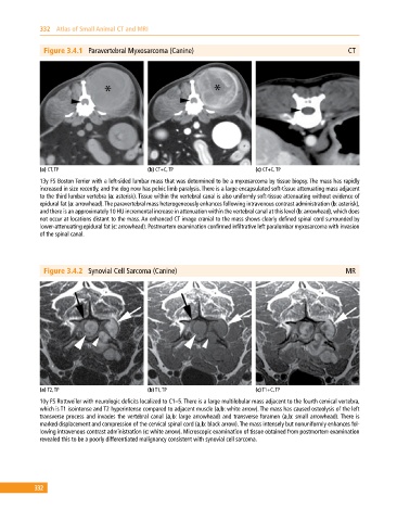
Figure 3.4.1 Paravertebral Myxosarcoma (Canine) CT
(a) CT, TP (b) CT+C, TP (c) CT+C, TP
13y FS Boston Terrier with a left‐sided lumbar mass that was determined to be a myxosarcoma by tissue biopsy. The mass has rapidly
increased in size recently, and the dog now has pelvic limb paralysis. There is a large encapsulated soft‐tissue attenuating mass adjacent
to the third lumbar vertebra (a: asterisk). Tissue within the vertebral canal is also uniformly soft‐tissue attenuating without evidence of
epidural fat (a: arrowhead). The paravertebral mass heterogeneously enhances following intravenous contrast administration (b: asterisk),
and there is an approximately 10 HU incremental increase in attenuation within the vertebral canal at this level (b: arrowhead), which does
not occur at locations distant to the mass. An enhanced CT image cranial to the mass shows clearly defined spinal cord surrounded by
lower‐attenuating epidural fat (c: arrowhead). Postmortem examination confirmed infiltrative left paralumbar myxosarcoma with invasion
of the spinal canal.
Figure 3.4.2 Synovial Cell Sarcoma (Canine) MR
(a) T2, TP (b) T1, TP (c) T1+C, TP
10y FS Rottweiler with neurologic deficits localized to C1–5. There is a large multilobular mass adjacent to the fourth cervical vertebra,
which is T1 isointense and T2 hyperintense compared to adjacent muscle (a,b: white arrow). The mass has caused osteolysis of the left
transverse process and invades the vertebral canal (a,b: large arrowhead) and transverse foramen (a,b: small arrowhead). There is
marked displacement and compression of the cervical spinal cord (a,b: black arrow). The mass intensely but nonuniformly enhances fol
lowing intravenous contrast administration (c: white arrow). Microscopic examination of tissue obtained from postmortem examination
revealed this to be a poorly differentiated malignancy consistent with synovial cell sarcoma.
332