
Page 345 - Atlas of Small Animal CT and MRI
P. 345
Neoplasia 335
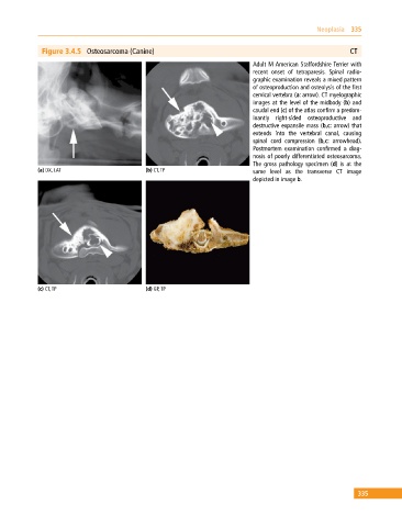
Figure 3.4.5 Osteosarcoma (Canine) CT
Adult M American Staffordshire Terrier with
recent onset of tetraparesis. Spinal radio
graphic examination reveals a mixed pattern
of osteoproduction and osteolysis of the first
cervical vertebra (a: arrow). CT myelographic
images at the level of the midbody (b) and
caudal end (c) of the atlas confirm a predo m
inantly right‐sided osteoproductive and
destructive expansile mass (b,c: arrow) that
extends into the vertebral canal, causing
spinal cord compression (b,c: arrowhead).
Postmortem examination confirmed a diag
nosis of poorly differentiated osteosarcoma.
The gross pathology specimen (d) is at the
(a) DX, LAT (b) CT, TP same level as the transverse CT image
depicted in image b.
(c) CT, TP (d) GP, TP
335