
Page 350 - Atlas of Small Animal CT and MRI
P. 350
340 Atlas of Small Animal CT and MRI
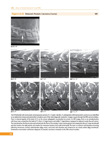
Figure 3.4.10 Metastatic Prostatic Carcinoma (Canine) MR
(a) DX, LAT
(b) T2, SP (c) T1, SP (d) T1+C, SP
(e) T2, TP (f) T1, TP (g) T1+C, TP
10y M Rottweiler with cervical pain and progressive paresis of 3–4 weeks’ duration. A cytologically confirmed prostatic carcinoma was identified
on an abdominal ultrasound examination included as part of the initial diagnostic evaluation. Images e–g are through the fifth cervical vertebra.
There is reduced opacity of the body of C5 on the survey radiographic examination (a: arrow). On MR images, there is an osteodestructive
soft‐tissue mass arising from the body of C5 that is T2 hyperintense and mildly T1 hyperintense compared to adjacent muscle (b,c,e,f: arrow).
The mass breaches the dorsal cortex and extends into the floor of the vertebral canal, causing spinal cord compression (b,c,e,f: large arrowhead)
and also involves the right pedicle and invades the transverse foramen (e,f: small arrowhead). The mass is uniformly and intensely enhancing
following intravenous contrast administration (d,g: arrow), and spinal cord elevation and compression are clearly evident (d,g: arrowhead).
Postmortem examination confirmed a diagnosis of prostatic carcinoma metastatic to the fifth cervical vertebra.
340