Page 354 - Atlas of Small Animal CT and MRI
P. 354

344  Atlas of Small Animal CT and MRI


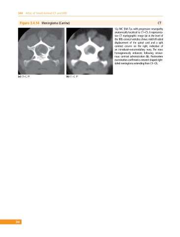
              Figure 3.4.14  Meningioma (Canine)                                                           CT
                                                                              12y MC Shih Tzu with progressive neuropathy
                                                                              anatomically localized to C1–C5. A representa­
                                                                              tive CT myelographic image (a) at the level of
                                                                              the fifth cervical vertebra shows mild left‐sided
                                                                                displacement of the spinal cord and a split
                                                                                contrast column on the right, indicative of
                                                                              an  intradural–extramedullary mass. The mass
                                                                              homogeneously enhances following intrave­
                                                                              nous  contrast  administration  (b).  Postmortem
                                                                              examination confirmed a crescent‐shaped right‐
                                                                              sided meningioma extending from C4–C6.


             (a) CT+C, TP                     (b) CT+C, TP


           344
   349   350   351   352   353   354   355   356   357   358   359