
Page 359 - Atlas of Small Animal CT and MRI
P. 359
Neoplasia 349
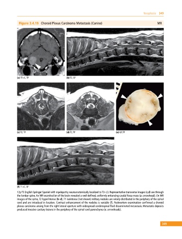
Figure 3.4.19 Choroid Plexus Carcinoma Metastasis (Canine) MR
(a) T1+C, TP (b) T2, SP
(c) T2, TP (d) T2, TP (e) GP, TP
(f) T1+C, SP
12y FS English Springer Spaniel with myelopathy neuroanatomically localized to T3–L3. Representative transverse images (c,d) are through
the lumbar spine. An MR examination of the brain revealed a well‐defined, uniformly enhancing caudal fossa mass (a: arrowhead). On MR
images of the spine, T2 hyperintense (b–d), T1 isointense (not shown) military nodules are widely distributed in the periphery of the spinal
cord and are intradural in location. Contrast enhancement of the nodules is variable (f). Postmortem examination confirmed a choroid
plexus carcinoma arising from the right lateral aperture with widespread cerebrospinal fluid disseminated metastasis. Metastatic deposits
produced invasive cavitary lesions in the periphery of the spinal cord parenchyma (e: arrowheads).
349