
Page 363 - Atlas of Small Animal CT and MRI
P. 363
Neoplasia 353
Figure 3.4.22 (Continued ) MR
(g) GP, DORS
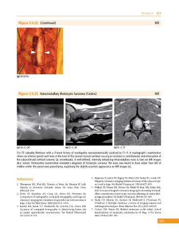
Figure 3.4.23 Intramedullary Histiocytic Sarcoma (Canine) MR
(a) DX+C, LAT (b) T1+C, SP (c) T1+C, TP
11y FS Labrador Retriever with a 3‐week history of myelopathy neuroanatomically localized to C1–5. A myelographic examination
shows an intrinsic spinal cord mass at the level of the second cervical vertebra causing an increase in cord diameter and attenuation of
the subarachnoid contrast columns (a: arrowheads). A well‐defined, intensely enhancing intramedullary mass is seen on MR images
(b,c: arrow). Postmortem examination revealed a diagnosis of histiocytic sarcoma. The mass was found to have arisen from left of
midline within the spinal cord parenchyma, explaining the slightly eccentric appearance on MR images (c).
References 4. Kippenes H, Gavin PR, Bagley RS, Silver GM, Tucker RL, Sande RD.
Magnetic resonance imaging features of tumors of the spine and spi
1. Thompson KG, Pool RR. Tumors of Bone. In: Meuten DJ (ed): nal cord in dogs. Vet Radiol Ultrasound. 1999;40:627–633.
Tumors in Domestic Animals. Ames, IA: Iowa State Press, 5. Wallack ST, Wisner ER, Werner JA, Walsh PJ, Kent MS, Fairley RA,
2002;248–255. et al. Accuracy of magnetic resonance imaging for estimating intramed
2. Davis GJ, Kapatkin AS, Craig LE, Heins GS, Wortman JA. ullary osteosarcoma extent in pre‐operative planning of canine limb‐
Comparison of radiography, computed tomography, and magnetic salvage procedures. Vet Radiol Ultrasound. 2002;43:432–441.
resonance imaging for evaluation of appendicular osteosarcoma in 6. Healy CF, Murray JG, Eustace SJ, Madewell J, O’Gorman PJ,
dogs. J Am Vet Med Assoc. 2002;220:1171–1176. O’Sullivan P. Multiple myeloma: a review of imaging features and
3. Karnik KS, Samii VF, Weisbrode SE, London CA, Green EM. radiological techniques. Bone Marrow Res. 2011;2011:583439.
Accuracy of computed tomography in determining lesion size 7. Cooley DM, Waters DJ. Skeletal metastasis as the initial clinical
in canine appendicular osteosarcoma. Vet Radiol Ultrasound. manifestation of metastatic carcinoma in 19 dogs. J Vet Intern
2012;53:273–279. Med. 1998;12:288–293.
353