
Page 361 - Atlas of Small Animal CT and MRI
P. 361
Neoplasia 351
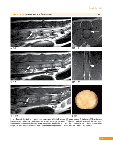
Figure 3.4.21 Glioblastoma Multiforme (Feline) MR
(a) T2, SP (b) T1+C, TP
(c) T1, SP (d) T1+C, DP
(e) T1+C, SP (f) GP, TP
9y MC Domestic Shorthair with recent‐onset progressive pelvic limb paresis. MR images show a T1 isointense, T2 hyperintense,
heterogeneously enhancing, ovoid intrinsic spinal cord mass at the level of the fifth lumbar vertebra (a–e: arrow). The gross speci
men (f) shows that the mass displaces neural parenchyma peripherally, resulting in the focal increase in cord diameter seen on MR
images (d). Microscopic examination confirmed a diagnosis of glioblastoma multiforme (WHO grade IV astrocytoma).
351