
Page 356 - Atlas of Small Animal CT and MRI
P. 356
346 Atlas of Small Animal CT and MRI
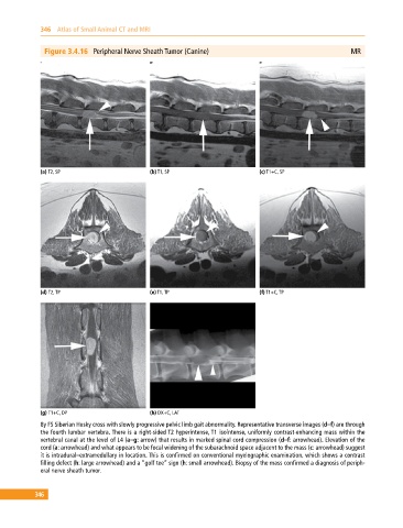
Figure 3.4.16 Peripheral Nerve Sheath Tumor (Canine) MR
(a) T2, SP (b) T1, SP (c) T1+C, SP
(d) T2, TP (e) T1, TP (f) T1+C, TP
(g) T1+C, DP (h) DX+C, LAT
8y FS Siberian Husky cross with slowly progressive pelvic limb gait abnormality. Representative transverse images (d–f) are through
the fourth lumbar vertebra. There is a right‐sided T2 hyperintense, T1 isointense, uniformly contrast‐enhancing mass within the
vertebral canal at the level of L4 (a–g: arrow) that results in marked spinal cord compression (d–f: arrowhead). Elevation of the
cord (a: arrowhead) and what appears to be focal widening of the subarachnoid space adjacent to the mass (c: arrowhead) suggest
it is intradural–extramedullary in location. This is confirmed on conventional myelographic examination, which shows a contrast
filling defect (h: large arrowhead) and a “golf tee” sign (h: small arrowhead). Biopsy of the mass confirmed a diagnosis of periph
eral nerve sheath tumor.
346