
Page 360 - Atlas of Small Animal CT and MRI
P. 360
350 Atlas of Small Animal CT and MRI
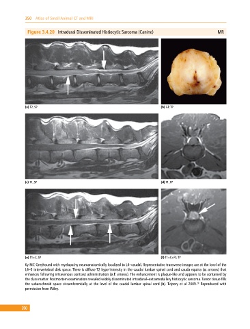
Figure 3.4.20 Intradural Disseminated Histiocytic Sarcoma (Canine) MR
(a) T2, SP (b) GP, TP
(c) T1, SP (d) T1, TP
(e) T1+C, SP (f) T1+C+FS, TP
6y MC Greyhound with myelopathy neuroanatomically localized to L4–caudal. Representative transverse images are at the level of the
L4–5 intervertebral disk space. There is diffuse T2 hyperintensity in the caudal lumbar spinal cord and cauda equina (a: arrows) that
enhances following intravenous contrast administration (e,f: arrows). The enhancement is plaque‐like and appears to be contained by
the dura matter. Postmortem examination revealed widely disseminated intradural–extramedullary histiocytic sarcoma. Tumor tissue fills
29
the subarachnoid space circumferentially at the level of the caudal lumbar spinal cord (b). Tzipory et al 2009. Reproduced with
permission from Wiley.
350