
Page 355 - Atlas of Small Animal CT and MRI
P. 355
Neoplasia 345
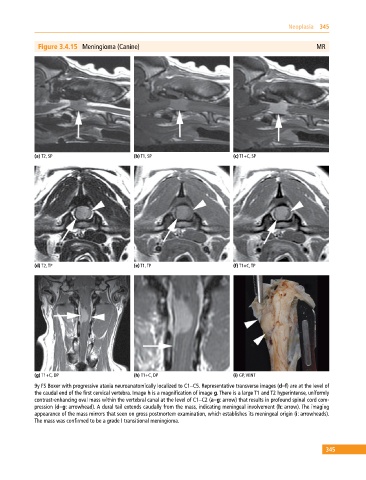
Figure 3.4.15 Meningioma (Canine) MR
(a) T2, SP (b) T1, SP (c) T1+C, SP
(d) T2, TP (e) T1, TP (f) T1+C, TP
(g) T1+C, DP (h) T1+C, DP (i) GP, VENT
9y FS Boxer with progressive ataxia neuroanatomically localized to C1–C5. Representative transverse images (d–f) are at the level of
the caudal end of the first cervical vertebra. Image h is a magnification of image g. There is a large T1 and T2 hyperintense, uniformly
contrast‐enhancing oval mass within the vertebral canal at the level of C1–C2 (a–g: arrow) that results in profound spinal cord com
pression (d–g: arrowhead). A dural tail extends caudally from the mass, indicating meningeal involvement (h: arrow). The imaging
appearance of the mass mirrors that seen on gross postmortem examination, which establishes its meningeal origin (i: arrowheads).
The mass was confirmed to be a grade I transitional meningioma.
345