
Page 351 - Atlas of Small Animal CT and MRI
P. 351
Neoplasia 341
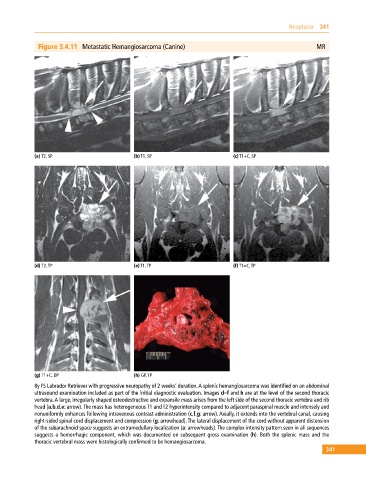
Figure 3.4.11 Metastatic Hemangiosarcoma (Canine) MR
(a) T2, SP (b) T1, SP (c) T1+C, SP
(d) T2, TP (e) T1, TP (f) T1+C, TP
(g) T1+C, DP (h) GP, TP
8y FS Labrador Retriever with progressive neuropathy of 2 weeks’ duration. A splenic hemangiosarcoma was identified on an abdominal
ultrasound examination included as part of the initial diagnostic evaluation. Images d–f and h are at the level of the second thoracic
vertebra. A large, irregularly shaped osteodestructive and expansile mass arises from the left side of the second thoracic vertebra and rib
head (a,b,d,e: arrow). The mass has heterogeneous T1 and T2 hyperintensity compared to adjacent paraspinal muscle and intensely and
nonuniformly enhances following intravenous contrast administration (c,f,g: arrow). Axially, it extends into the vertebral canal, causing
right‐sided spinal cord displacement and compression (g: arrowhead). The lateral displacement of the cord without apparent distension
of the subarachnoid space suggests an extramedullary localization (a: arrowheads). The complex intensity pattern seen in all sequences
suggests a hemorrhagic component, which was documented on subsequent gross examination (h). Both the splenic mass and the
thoracic vertebral mass were histologically confirmed to be hemangiosarcoma.
341