
Page 347 - Atlas of Small Animal CT and MRI
P. 347
Neoplasia 337
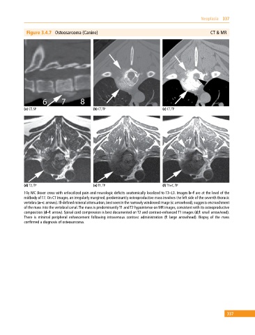
Figure 3.4.7 Osteosarcoma (Canine) CT & MR
(a) CT, SP (b) CT, TP (c) CT, TP
(d) T2, TP (e) T1, TP (f) T1+C, TP
10y MC Boxer cross with unlocalized pain and neurologic deficits anatomically localized to T3–L3. Images b–f are at the level of the
midbody of T7. On CT images, an irregularly margined, predominantly osteoproductive mass involves the left side of the seventh thoracic
vertebra (a–c: arrows). Ill‐defined mineral attenuation, best seen in the narrowly windowed image (c: arrowhead), suggests encroachment
of the mass into the vertebral canal. The mass is predominantly T1 and T2 hypointense on MR images, consistent with its osteoproductive
composition (d–f: arrow). Spinal cord compression is best documented on T2 and contrast‐enhanced T1 images (d,f: small arrowhead).
There is minimal peripheral enhancement following intravenous contrast administration (f: large arrowhead). Biopsy of the mass
confirmed a diagnosis of osteosarcoma.
337