
Page 352 - Atlas of Small Animal CT and MRI
P. 352
342 Atlas of Small Animal CT and MRI
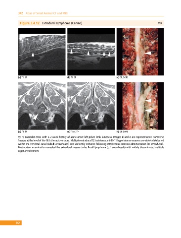
Figure 3.4.12 Extradural Lymphoma (Canine) MR
(a) T2, SP (b) T2, SP (c) GP, DORS
(d) T1, TP (e) T1+C, TP (f) GP, DORS
9y FS Labrador cross with a 2‐week history of acute‐onset left pelvic limb lameness. Images d and e are representative transverse
images at the level of the fifth thoracic vertebra. Multiple extradural T2 isointense, mildly T1 hyperintense masses are widely distributed
within the vertebral canal (a,b,d: arrowheads) and uniformly enhance following intravenous contrast administration (e: arrowhead).
Postmortem examination revealed the extradural masses to be B‐cell lymphoma (c,f: arrowheads) with widely disseminated multiple
organ involvement.
342