
Page 357 - Atlas of Small Animal CT and MRI
P. 357
Neoplasia 347
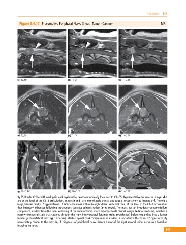
Figure 3.4.17 Presumptive Peripheral Nerve Sheath Tumor (Canine) MR
(a) T2, SP (b) T1, SP (c) T1+C, SP
(d) T2, TP (e) T1, TP (f) T1+C, TP
(g) T1+C, DP (h) T1+C, TP (i) T1+C, TP
9y FS Border Collie with neck pain and myelopathy neuroanatomically localized to C1–C5. Representative transverse images d–f
are at the level of the C1–2 articulation. Images h and i are immediately cranial and caudal, respectively, to images d–f. There is a
large, lobular, mildly T2 hyperintense, T1 isointense mass within the right dorsal vertebral canal at the level of the C1–2 articulation
that intensely enhances following intravenous contrast administration (a–h: arrow). The mass has an intradural–extramedullary
component, evident from the focal widening of the subarachnoid space adjacent to its caudal margin (a,b: arrowhead), and has a
narrow extradural stalk that courses through the right intervertebral foramen (g,h: arrowheads) before expanding into a larger,
lobular juxtavertebral mass (g,i: asterisk). Marked spinal cord compression is evident, associated with central T2 hyperintensity
immediately caudal to the mass (a). A diagnosis of peripheral nerve sheath tumor of the right second spinal nerve was based on
imaging features.
347