
Page 362 - Atlas of Small Animal CT and MRI
P. 362
352 Atlas of Small Animal CT and MRI
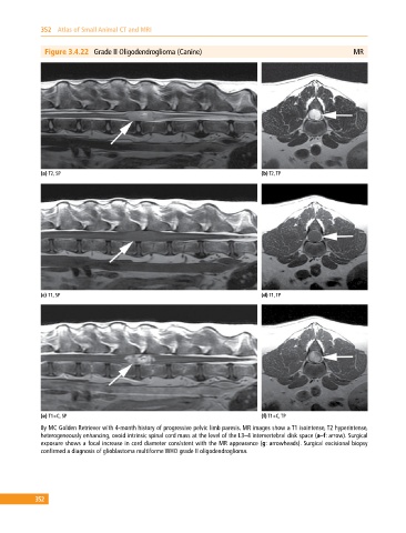
Figure 3.4.22 Grade II Oligodendroglioma (Canine) MR
(a) T2, SP (b) T2, TP
(c) T1, SP (d) T1, TP
(e) T1+C, SP (f) T1+C, TP
8y MC Golden Retriever with 4‐month history of progressive pelvic limb paresis. MR images show a T1 isointense, T2 hyperintense,
heterogeneously enhancing, ovoid intrinsic spinal cord mass at the level of the L3–4 intervertebral disk space (a–f: arrow). Surgical
exposure shows a focal increase in cord diameter consistent with the MR appearance (g: arrowheads). Surgical excisional biopsy
confirmed a diagnosis of glioblastoma multiforme WHO grade II oligodendroglioma.
352