
Page 358 - Atlas of Small Animal CT and MRI
P. 358
348 Atlas of Small Animal CT and MRI
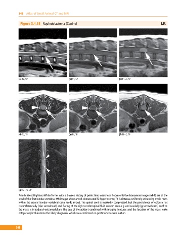
Figure 3.4.18 Nephroblastoma (Canine) MR
(a) T2, SP (b) T1, SP (c) T1+C, SP
(d) T2, TP (e) T1, TP (f) T1+C, TP
(g) T2+FS, DP
7mo M West Highland White Terrier with a 2‐week history of pelvic limb weakness. Representative transverse images (d–f) are at the
level of the first lumbar vertebra. MR images show a well‐demarcated T2 hyperintense, T1 isointense, uniformly enhancing ovoid mass
within the cranial lumbar vertebral canal (a–f: arrow). The spinal cord is markedly compressed, but the persistence of epidural fat
circumferentially (d,e: arrowhead) and flaring of the right cerebrospinal fluid column cranially and caudally (g: arrowheads) confirm
the mass is intradural–extramedullary. The age of the patient combined with imaging features and the location of the mass make
ectopic nephroblastoma the likely diagnosis, which was confirmed on postmortem examination.
348