
Page 343 - Atlas of Small Animal CT and MRI
P. 343
Neoplasia 333
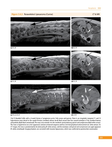
Figure 3.4.3 Paravertebral Liposarcoma (Canine) CT & MR
(a) T2, SP (b) T2, TP
(c) T1, SP (d) T1, TP
(e) T1+C+FS, SP (f) CT, TP
14y FS Bearded Collie with a 3‐week history of progressive pelvic limb ataxia and paresis. There is an irregularly margined T1 and T2
hyperintense mass dorsal to the caudal thoracic vertebral column (a–d: black arrow) that has caused osteolysis of the vertebral lamina
and pedicles (b,d: black arrowhead). The mass also extends into the vertebral canal producing spinal cord compression (b,d: white arrow).
The mass is hypointense on a fat‐suppressed contrast‐enhanced sequence and has minimal peripheral enhancement (e: white arrow). On
CT images, the mass is predominantly fat attenuating (f: white arrow), and the osteolysis and spinal cord compression are again apparent
(f: white arrowhead). Imaging features are consistent with invasive liposarcoma, which was confirmed on postmortem examination.
333