
Page 382 - Atlas of Small Animal CT and MRI
P. 382
372 Atlas of Small Animal CT and MRI
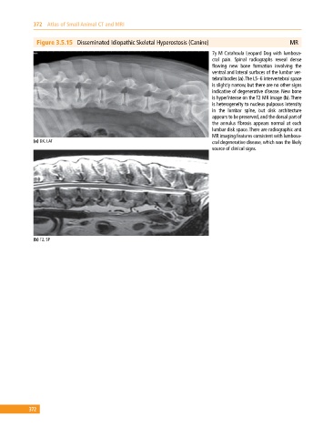
Figure 3.5.15 Disseminated Idiopathic Skeletal Hyperostosis (Canine) MR
7y M Catahoula Leopard Dog with lumbosa
cral pain. Spinal radiographs reveal dense
flowing new bone formation involving the
ventral and lateral surfaces of the lumbar ver
tebral bodies (a). The L5–6 intervertebral space
is slightly narrow, but there are no other signs
indicative of degenerative disease. New bone
is hyperintense on the T2 MR image (b). There
is heterogeneity to nucleus pulposus intensity
in the lumbar spine, but disk architecture
appears to be preserved, and the dorsal part of
the annulus fibrosis appears normal at each
lumbar disk space. There are radiographic and
MR imaging features consistent with lumbosa
(a) DX, LAT cral degenerative disease, which was the likely
source of clinical signs.
(b) T2, SP
372