
Page 379 - Atlas of Small Animal CT and MRI
P. 379
Intervertebral disk disease and other degenerative disorders 369
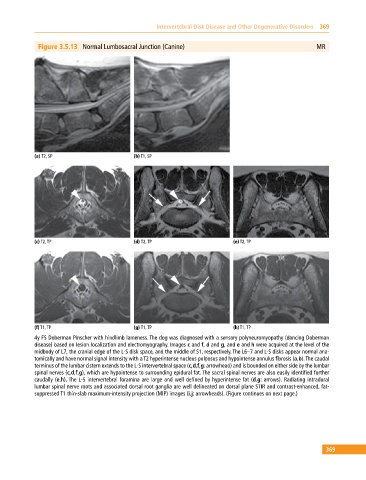
Figure 3.5.13 Normal Lumbosacral Junction (Canine) MR
(a) T2, SP (b) T1, SP
(c) T2, TP (d) T2, TP (e) T2, TP
(f) T1, TP (g) T1, TP (h) T1, TP
4y FS Doberman Pinscher with hindlimb lameness. The dog was diagnosed with a sensory polyneuromyopathy (dancing Doberman
disease) based on lesion localization and electromyography. Images c and f, d and g, and e and h were acquired at the level of the
midbody of L7, the cranial edge of the L‐S disk space, and the middle of S1, respectively. The L6–7 and L‐S disks appear normal ana
tomically and have normal signal intensity with a T2 hyperintense nucleus pulposus and hypointense annulus fibrosis (a,b). The caudal
terminus of the lumbar cistern extends to the L‐S intervertebral space (c,d,f,g: arrowhead) and is bounded on either side by the lumbar
spinal nerves (c,d,f,g), which are hypointense to surrounding epidural fat. The sacral spinal nerves are also easily identified further
caudally (e,h). The L‐S intervertebral foramina are large and well defined by hyperintense fat (d,g: arrows). Radiating intradural
lumbar spinal nerve roots and associated dorsal root ganglia are well delineated on dorsal plane STIR and contrast‐enhanced, fat‐
suppressed T1 thin‐slab maximum‐intensity projection (MIP) images (i,j: arrowheads). (Figure continues on next page.)
369