
Page 374 - Atlas of Small Animal CT and MRI
P. 374
364 Atlas of Small Animal CT and MRI
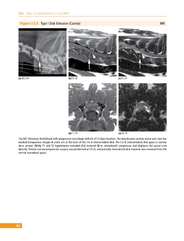
Figure 3.5.8 Type I Disk Extrusion (Canine) MR
(a) DX, LAT (b) T1, SP (c) T2, SP
(d) T1, TP (e) T2, TP
13y MC Miniature Dachshund with progressive neurologic deficits of 10 days duration. The dog became acutely worse and now has
marked tetraparesis. Images d and e are at the level of the C5–6 intervertebral disk. The C5–6 intervertebral disk space is narrow
(a–c: arrow). Mildly T1 and T2 hypointense extruded disk material (b–e: arrowhead) compresses and displaces the spinal cord
dorsally. Ventral slot decompression surgery was performed at C5–6, and partially mineralized disk material was removed from the
ventral extradural space.
364