
Page 376 - Atlas of Small Animal CT and MRI
P. 376
366 Atlas of Small Animal CT and MRI
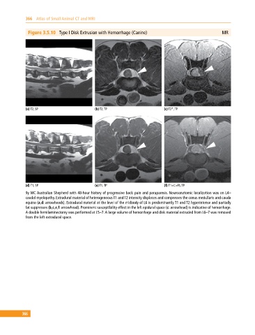
Figure 3.5.10 Type I Disk Extrusion with Hemorrhage (Canine) MR
(a) T2, SP (b) T2, TP (c) T2*, TP
(d) T1, SP (e) T1, TP (f) T1+C+FS, TP
9y MC Australian Shepherd with 48‐hour history of progressive back pain and paraparesis. Neuroanatomic localization was an L4–
caudal myelopathy. Extradural material of heterogeneous T1 and T2 intensity displaces and compresses the conus medullaris and cauda
equina (a,d: arrowheads). Extradural material at the level of the midbody of L6 is predominantly T1 and T2 hyperintense and partially
fat suppresses (b,c,e,f: arrowhead). Prominent susceptibility effect in the left epidural space (c: arrowhead) is indicative of hemorrhage.
A double hemilaminectomy was performed at L5–7. A large volume of hemorrhage and disk material extruded from L6–7 was removed
from the left extradural space.
366