
Page 373 - Atlas of Small Animal CT and MRI
P. 373
Intervertebral disk disease and other degenerative disorders 363
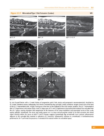
Figure 3.5.7 Mineralized Type I Disk Extrusion (Canine) MR
(a) T2, SP (b) T1, SP (c) T1+C+FS, SP
(d) T2, TP (e) T1, TP
(f) T2, TP (g) T1, TP
5y Jack Russell Terrier with a 2‐week history of progressive pelvic limb ataxia and paraparesis neuroanatomically localized to
L6–caudal. Vertebral column radiographs (not shown) revealed the dog had eight lumbar vertebrae. Images d and e are at the level
of the L6–7 intervertebral disk. Images f and g are at the level of the midbody of the sixth lumbar vertebra. The L6–7 intervertebral
disk is T2 hypointense and narrow (a,b: arrow). A large mass of T1 and T2 hypointense mineralized disk material has extruded from
the L6–7 space, causing elevation and compression of the terminus of the conus medullaris and associated nerves of the cauda
equina (a,b,d,e: arrowhead). The conus and cauda equina are in a more normal position cranial to the disk extrusion, but mixed
signal intensity within the conus on both T1 and T2 images suggests hemorrhage (f,g: arrowhead). Local contrast enhancement
adjacent to the extruded disk material is indicative of a traumatic inflammatory response (c: arrowhead). A hemilaminectomy
performed at L6–7 confirmed the presence of extruded disk material within the extradural space.
363