
Page 370 - Atlas of Small Animal CT and MRI
P. 370
360 Atlas of Small Animal CT and MRI
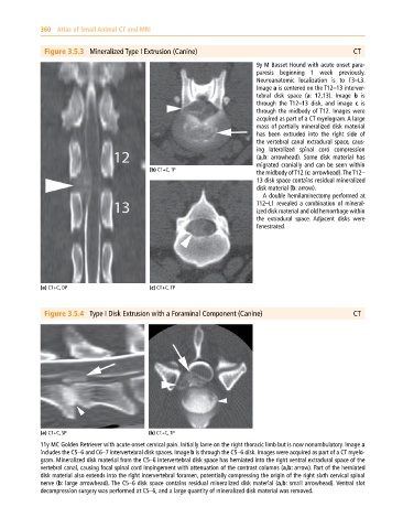
Figure 3.5.3 Mineralized Type I Extrusion (Canine) CT
9y M Basset Hound with acute onset para
paresis beginning 1 week previously.
Neuroanatomic localization is to T3–L3.
Image a is centered on the T12–13 interver
tebral disk space (a: 12,13). Image b is
through the T12–13 disk, and image c is
through the midbody of T12. Images were
acquired as part of a CT myelogram. A large
mass of partially mineralized disk material
has been extruded into the right side of
the vertebral canal extradural space, caus
ing lateralized spinal cord compression
(a,b: arrowhead). Some disk material has
migrated cranially and can be seen within
(b) CT+C, TP
the midbody of T12 (c: arrowhead). The T12–
13 disk space contains residual mineralized
disk material (b: arrow).
A double hemilaminectomy performed at
T12–L1 revealed a combination of mineral
ized disk material and old hemorrhage within
the extradural space. Adjacent disks were
fenestrated.
(a) CT+C, DP (c) CT+C, TP
Figure 3.5.4 Type I Disk Extrusion with a Foraminal Component (Canine) CT
(a) CT+C, SP (b) CT+C, TP
11y MC Golden Retriever with acute‐onset cervical pain. Initially lame on the right thoracic limb but is now nonambulatory. Image a
includes the C5–6 and C6–7 intervertebral disk spaces. Image b is through the C5–6 disk. Images were acquired as part of a CT myelo
gram. Mineralized disk material from the C5–6 intervertebral disk space has herniated into the right ventral extradural space of the
vertebral canal, causing focal spinal cord impingement with attenuation of the contrast columns (a,b: arrow). Part of the herniated
disk material also extends into the right intervertebral foramen, potentially compressing the origin of the right sixth cervical spinal
nerve (b: large arrowhead). The C5–6 disk space contains residual mineralized disk material (a,b: small arrowhead). Ventral slot
decompression surgery was performed at C5–6, and a large quantity of mineralized disk material was removed.