
Page 372 - Atlas of Small Animal CT and MRI
P. 372
362 Atlas of Small Animal CT and MRI
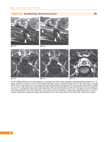
Figure 3.5.6 Mineralized Type I Disk Extrusion (Canine) MR
(a) T1, SP (b) T2, SP
(c) T1, TP (d) T2, TP (e) T2, TP
10y MC Alaskan Malamute cross with progressive cervical pain and recent ataxia and paresis neuroanatomically localized to C1–C5.
Images a and b are centered on the C4–5 intervertebral disk space. Images c and d are through the C4–5 disk, and image e is through
the C3–4 disk. A large volume of T1 and T2 hypointense disk material has been extruded into the ventral extradural space of the vertebral
canal at C4–5, causing dorsal displacement and compression of the spinal cord (a–d: arrow). The C4–5 disk space is narrow and hypoin
tense (a,b: arrowhead). The normal appearance of the spinal cord and surrounding T2 hyperintense subarachnoid space is shown in
image e. Ventral slot decompression surgery was performed at C4–5, and a large volume of mineralized disk material was removed.
362