
Page 377 - Atlas of Small Animal CT and MRI
P. 377
Intervertebral disk disease and other degenerative disorders 367
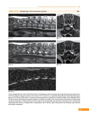
Figure 3.5.11 Multiple Type II Disk Protrusions (Canine) MR
(a) T1, SP (b) T2, TP
(c) T2, SP (d) T2, TP
(e) SSTSE, SP
13y MC Labrador Retriever with 4‐week history of pelvic limb paraparesis with acute progression of signs. Neuroanatomic localization is
to T3–L3 myelopathy. Images b and d are at the level of T12–13 and T13–L1, respectively. Thoracolumbar intervertebral disk spaces
appear to be of normal width and are T1 isointense and heterogeneously T2 hyperintense, indicating variable nuclear dehydration (a,c).
The dorsal annulus fibrosis/dorsal longitudinal ligament is prominent at multiple levels, elevating and compressing the overlying spinal
cord (b–d: arrowheads). A single‐shot turbo spin‐echo image of the thoracolumbar spine documents discontinuity of the subarachnoid
cerebrospinal fluid columns at multiple levels, corresponding to sites of Hansen’s type II disk protrusion (e). Neurologic signs improved
with medical management.
367