
Page 378 - Atlas of Small Animal CT and MRI
P. 378
368 Atlas of Small Animal CT and MRI
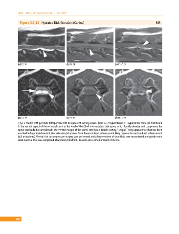
Figure 3.5.12 Hydrated Disk Extrusion (Canine) MR
(a) T2, SP (b) T1, SP (c) T1+C, SP
(d) T2, TP (e) T1, TP (f) T1+C, TP
10y FS Poodle with peracute tetraparesis with no apparent inciting cause. There is T2 hyperintense, T1 hypointense material distributed
in the ventral aspect of the vertebral canal at the level of the C3–4 intervertebral disk space, which focally elevates and compresses the
spinal cord (a,b,d,e: arrowhead). The ventral margin of the spinal cord has a double arching “seagull” wing appearance that has been
ascribed to high liquid content disk extrusion (d: arrow). Focal linear contrast enhancement likely represents reactive dural enhancement
(c,f: arrowhead). Ventral slot decompression surgery was performed and a large volume of clear fluid was encountered along with more
solid material that was composed of atypical chondroid‐like cells and a small amount of matrix.
368