
Page 383 - Atlas of Small Animal CT and MRI
P. 383
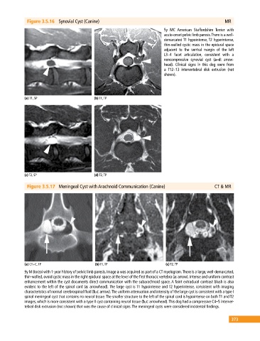
Figure 3.5.16 Synovial Cyst (Canine) MR
5y MC American Staffordshire Terrier with
acute‐onset pelvic limb paresis. There is a well‐
demarcated T1 hypointense, T2 hyperintense,
thin‐walled cystic mass in the epidural space
adjacent to the ventral margin of the left
L3–4 facet articulation, consistent with a
noncompressive synovial cyst (a–d: arrow
head). Clinical signs in this dog were from
a T12–13 intervertebral disk extrusion (not
shown).
(a) T1, SP (b) T1, TP
(c) T2, SP (d) T2, TP
Figure 3.5.17 Meningeal Cyst with Arachnoid Communication (Canine) CT & MR
(a) CT+C, TP (b) T1, TP (c) T2, TP
9y M Borzoi with 1‐year history of pelvic limb paresis. Image a was acquired as part of a CT myelogram. There is a large, well‐demarcated,
thin‐walled, ovoid cystic mass in the right epidural space at the level of the first thoracic vertebra (a: arrow). Intense and uniform contrast
enhancement within the cyst documents direct communication with the subarachnoid space. A faint extradural contrast blush is also
evident to the left of the spinal cord (a: arrowhead). The large cyst is T1 hypointense and T2 hyperintense, consistent with imaging
characteristics of normal cerebrospinal fluid (b,c: arrow). The uniform attenuation and intensity of the large cyst is consistent with a type I
spinal meningeal cyst that contains no neural tissue. The smaller structure to the left of the spinal cord is hypointense on both T1 and T2
images, which is more consistent with a type II cyst containing neural tissue (b,c: arrowhead). This dog had a compressive C4–5 interver
tebral disk extrusion (not shown) that was the cause of clinical signs. The meningeal cysts were considered incidental findings.
373