
Page 388 - Atlas of Small Animal CT and MRI
P. 388
378 Atlas of Small Animal CT and MRI
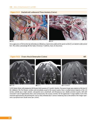
Figure 3.6.1 Brachial and Lumbosacral Plexus Anatomy (Canine)
(a) LLAT (b) LLAT
Gross appearance of the brachial (a) and lumbosacral (b) plexus. Spinal nerves (a,b) and the spinal cord (b: SC) are labeled in abbreviated
form. The authors acknowledge Mr. Ken Taylor, University of California, Davis, for dissections.
Figure 3.6.2 Chronic Muscle Denervation (Canine) MR
(a) T1, TP (b) T2, TP (c) T1+C+FS, TP
7y MC Boston Terrier with progressive left thoracic limb lameness of 4 months’ duration. Transverse images were acquired at the level of
the midbody of the first thoracic vertebra and immediately caudal to the scapular spines. There is marked volume reduction of the left
serratus ventralis (a–c: single asterisk), subscapularis (a–c: arrowhead), and infraspinatus (a–c: double asterisks) muscles, with mild
increased T1 and T2 hyperintensity that is most pronounced in the serratus ventralis. The fat‐suppressed T1 image nullifies much of the
increased signal intensity, documenting the cause as fatty infiltration (b). A contrast‐enhancing mass at the bottom of the images repre-
sents a peripheral nerve sheath tumor (a–c: arrow).
378