
Page 391 - Atlas of Small Animal CT and MRI
P. 391
Brachial and Lumbosacral Plexus 381
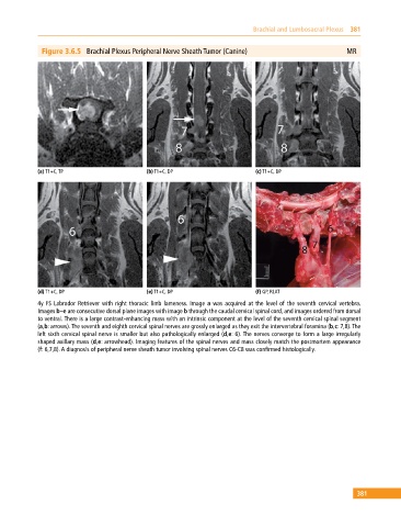
Figure 3.6.5 Brachial Plexus Peripheral Nerve Sheath Tumor (Canine) MR
(a) T1+C, TP (b) T1+C, DP (c) T1+C, DP
(d) T1+C, DP (e) T1+C, DP (f) GP, RLAT
4y FS Labrador Retriever with right thoracic limb lameness. Image a was acquired at the level of the seventh cervical vertebra.
Images b–e are consecutive dorsal plane images with image b through the caudal cervical spinal cord, and images ordered from dorsal
to ventral. There is a large contrast‐enhancing mass with an intrinsic component at the level of the seventh cervical spinal segment
(a,b: arrows). The seventh and eighth cervical spinal nerves are grossly enlarged as they exit the intervertebral foramina (b,c: 7,8). The
left sixth cervical spinal nerve is smaller but also pathologically enlarged (d,e: 6). The nerves converge to form a large irregularly
shaped axillary mass (d,e: arrowhead). Imaging features of the spinal nerves and mass closely match the postmortem appearance
(f: 6,7,8). A diagnosis of peripheral nerve sheath tumor involving spinal nerves C6‐C8 was confirmed histologically.
381