Page 396 - Atlas of Small Animal CT and MRI
P. 396
386  Atlas of Small Animal CT and MRI


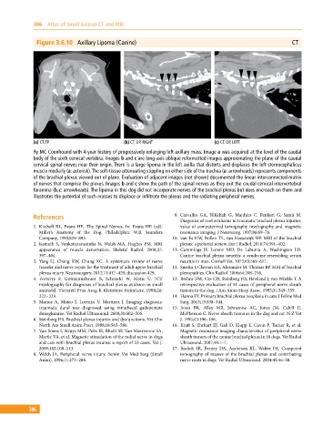
              Figure 3.6.10  Axillary Lipoma (Canine)                                                      CT


             (a) CT, TP                       (b) CT, OP, RIGHT                (c) CT, OP, LEFT
             9y MC Coonhound with 4‐year history of progressively enlarging left axillary mass. Image a was acquired at the level of the caudal
             body of the sixth cervical vertebra. Images b and c are long‐axis oblique reformatted images approximating the plane of the caudal
             cervical spinal nerves near their origin. There is a large lipoma in the left axilla that distorts and displaces the left sternocephalicus
             muscle medially (a: asterisk). The soft‐tissue attenuating stippling on either side of the trachea (a: arrowheads) represents components
             of the brachial plexus viewed out of plane. Evaluation of adjacent images (not shown) documented the linear interconnected matrix
             of nerves that comprise the plexus. Images b and c show the path of the spinal nerves as they exit the caudal cervical intervertebral
             foramina (b,c: arrowheads). The lipoma in this dog did not incorporate nerves of the brachial plexus but does encroach on them and
             illustrates the potential of such masses to displace or infiltrate the plexus and the radiating peripheral nerves.


             References                                         9.  Carvalho GA, Nikkhah G, Matthies C, Penkert G, Samii M.
                                                                  Diagnosis of root avulsions in traumatic brachial plexus injuries:
             1.  Kitchell  RL, Evans  HE. The Spinal Nerves. In: Evans HE  (ed):   value of computerized tomography myelography and magnetic
               Miller’s Anatomy of the Dog. Philadelphia: W.B. Saunders   resonance imaging. J Neurosurg. 1997;86:69–76.
               Company, 1993;829–893.                           10.  van Es HW, Bollen TL, van Heesewijk HP. MRI of the brachial
             2.  Kamath  S,  Venkatanarasimha  N,  Walsh  MA,  Hughes  PM.  MRI   plexus: a pictorial review. Eur J Radiol. 2010;74:391–402.
               appearance of muscle denervation. Skeletal Radiol. 2008;37:   11.  Cummings JF, Lorenz MD, De Lahunta A, Washington LD.
               397–404.                                           Canine brachial plexus neuritis: a syndrome resembling serum
             3.  Yang LJ, Chang KW, Chung KC. A systematic review of nerve   neuritis in man. Cornell Vet. 1973;63:589–617.
               transfer and nerve repair for the treatment of adult upper brachial   12.  Sureka J, Cherian RA, Alexander M, Thomas BP. MRI of brachial
               plexus injury. Neurosurgery. 2012;71:417–429; discussion 429.  plexopathies. Clin Radiol. 2009;64:208–218.
             4.  Forterre F, Gutmannsbauer B, Schmahl W, Matis U. [CT   13.  Brehm DM, Vite CH, Steinberg HS, Haviland J, van Winkle T. A
                 myelography for diagnosis of brachial plexus avulsion in small   retrospective evaluation of 51 cases of peripheral nerve sheath
               animals]. Tierarztl Prax Ausg K Kleintiere Heimtiere. 1998;26:   tumors in the dog. J Am Anim Hosp Assoc. 1995;31:349–359.
               322–329.                                         14.  Hanna FY. Primary brachial plexus neoplasia in cats. J Feline Med
             5.  Munoz A, Mateo I, Lorenzo V, Martinez J. Imaging diagnosis:   Surg. 2013;15:338–344.
                 traumatic dural tear diagnosed using intrathecal gadopentate   15.  Jones BR, Alley MR, Johnstone AC, Jones JM, Cahill JI,
               dimeglumine. Vet Radiol Ultrasound. 2009;50:502–505.  McPherson C. Nerve sheath tumours in the dog and cat. N Z Vet
             6.  Steinberg HS. Brachial plexus injuries and dysfunctions. Vet Clin   J. 1995;43:190–196.
               North Am Small Anim Pract. 1988;18:565–580.      16.  Kraft S, Ehrhart EJ, Gall D, Klopp L, Gavin P, Tucker R, et al.
             7.  Van Soens I, Struys MM, Polis IE, Bhatti SF, Van Meervenne SA,   Magnetic resonance imaging characteristics of peripheral nerve
               Martle VA, et al. Magnetic stimulation of the radial nerve in dogs   sheath tumors of the canine brachial plexus in 18 dogs. Vet Radiol
               and cats with brachial plexus trauma: a report of 53 cases. Vet J.   Ultrasound. 2007;48:1–7.
               2009;182:108–113.                                17.  Rudich SR, Feeney DA, Anderson KL, Walter PA. Computed
             8.  Welch  JA.  Peripheral nerve injury. Semin  Vet Med Surg  (Small   tomography of masses of the brachial plexus and contributing
               Anim). 1996;11:273–284.                            nerve roots in dogs. Vet Radiol Ultrasound. 2004;45:46–50.


           386

