
Page 393 - Atlas of Small Animal CT and MRI
P. 393
Brachial and Lumbosacral Plexus 383
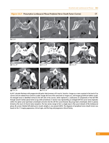
Figure 3.6.7 Presumptive Lumbosacral Plexus Peripheral Nerve Sheath Tumor (Canine) CT
(a) CT, TP (b) CT, TP (c) CT, TP
(d) CT+C, TP (e) CT+C, TP (f) CT+C, TP
(g) CT+C, TP (h) CT+C, TP
6y MC Labrador Retriever with progressive left pelvic limb lameness of 6 months’ duration. Images a–c were acquired at the level of the
sacrum and are ordered from cranial to caudal. Images d–f are at the same level as images a–c, and images g and h are further caudal.
An enlarged nerve is identified ventral to the left side of the sacrum, which represents a coalescence of components of the left fifth
through seventh lumbar spinal nerves (a–g: white arrowhead). A second mass representing an enlarged left first sacral nerve originates
within the spinal canal (a,d: black arrowhead) and exits into the left first sacral foramen (b,c,e–g: black arrowhead), which is grossly
dilated as the result of chronic bone resorption. The two nerves merge to form a single mass in the sacral network of the lumbosacral
plexus (h: arrow). Marked left‐sided pelvic muscle atrophy is also present (d–h). The diagnosis of peripheral nerve sheath tumor was
based on the CT imaging appearance, clinical signs, and the long and progressive clinical history.
383