
Page 390 - Atlas of Small Animal CT and MRI
P. 390
380 Atlas of Small Animal CT and MRI
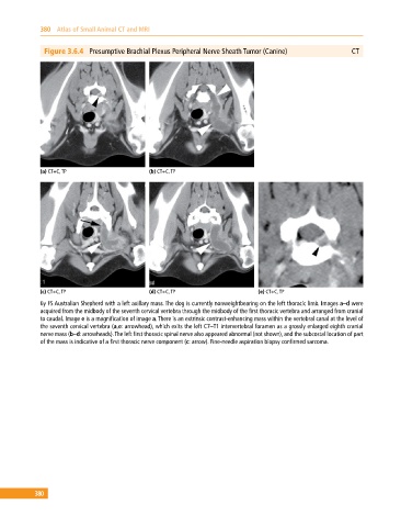
Figure 3.6.4 Presumptive Brachial Plexus Peripheral Nerve Sheath Tumor (Canine) CT
(a) CT+C, TP (b) CT+C, TP
(c) CT+C, TP (d) CT+C, TP (e) CT+C, TP
6y FS Australian Shepherd with a left axillary mass. The dog is currently nonweightbearing on the left thoracic limb. Images a–d were
acquired from the midbody of the seventh cervical vertebra through the midbody of the first thoracic vertebra and arranged from cranial
to caudal. Image e is a magnification of image a. There is an extrinsic contrast‐enhancing mass within the vertebral canal at the level of
the seventh cervical vertebra (a,e: arrowhead), which exits the left C7–T1 intervertebral foramen as a grossly enlarged eighth cranial
nerve mass (b–d: arrowheads). The left first thoracic spinal nerve also appeared abnormal (not shown), and the subcostal location of part
of the mass is indicative of a first thoracic nerve component (c: arrow). Fine‐needle aspiration biopsy confirmed sarcoma.
380