
Page 389 - Atlas of Small Animal CT and MRI
P. 389
Brachial and Lumbosacral Plexus 379
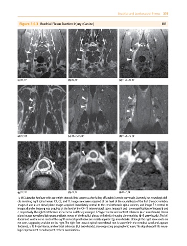
Figure 3.6.3 Brachial Plexus Traction Injury (Canine) MR
(a) T1, TP (b) T2, TP (c) T1+C+FS, TP
(d) T2, DP (e) T1+C+FS, DP (f) T1+C+FS, DP
(g) T2, TP (h) T2, TP (i) T1+C, TP
1y MC Labrador Retriever with acute right thoracic limb lameness after falling off a table 3 weeks previously. Currently has neurologic defi-
cits involving right spinal nerves C7, C8, and T1. Images a–c were acquired at the level of the caudal body of the first thoracic vertebra.
Images d and e are dorsal plane images acquired immediately ventral to the cervicothoracic spinal column, and image f is ventral to
images d and e. Image g was acquired at the level of the C7–T1 intervertebral space. Images h and i are magnifications of images b and
c, respectively. The right first thoracic spinal nerve is diffusely enlarged, T2 hyperintense and contrast enhances (a–c: arrowheads). Dorsal
plane images reveal multiple postganglionic nerves of the brachial plexus with similar imaging abnormalities (d–f: arrowheads). The left
dorsal and ventral nerve roots of the eighth cervical spinal nerve are readily apparent (g: arrowheads), although the right nerve roots are
not seen, suggesting avulsion on the right. The right first thoracic spinal nerve dorsal root is seen within the vertebral canal and appears
thickened, is T2 hyperintense, and contrast enhances (h,i: arrowheads), also suggesting preganglionic injury. The dog showed little neuro-
logic improvement on subsequent recheck examinations.