
Page 392 - Atlas of Small Animal CT and MRI
P. 392
382 Atlas of Small Animal CT and MRI
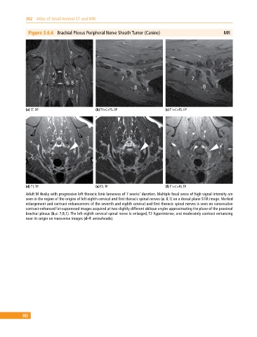
Figure 3.6.6 Brachial Plexus Peripheral Nerve Sheath Tumor (Canine) MR
(a) ST, DP (b) T1+C+FS, OP (c) T1+C+FS, OP
(d) T1, TP (e) T2, TP (f) T1+C+FS, TP
Adult M Husky with progressive left thoracic limb lameness of 7 weeks’ duration. Multiple focal areas of high signal intensity are
seen in the region of the origins of left eighth cervical and first thoracic spinal nerves (a: 8,1) on a dorsal plane STIR image. Marked
enlargement and contrast enhancement of the seventh and eighth cervical and first thoracic spinal nerves is seen on consecutive
contrast‐enhanced fat‐suppressed images acquired at two slightly different oblique angles approximating the plane of the proximal
brachial plexus (b,c: 7,8,1). The left eighth cervical spinal nerve is enlarged, T2 hyperintense, and moderately contrast enhancing
near its origin on transverse images (d–f: arrowheads).
382